Primary topic: AI in Diagnostic Imaging Centers (X-Ray, MRI, CT)
Research focus: Artificial intelligence in X-ray, computed tomography, magnetic resonance imaging, radiology workflow automation, image reconstruction, AI-assisted detection, triage, segmentation, quantitative imaging, reporting, PACS/RIS integration, quality assurance, predictive analytics, patient communication, and AI governance.
AI in Diagnostic Imaging Centers
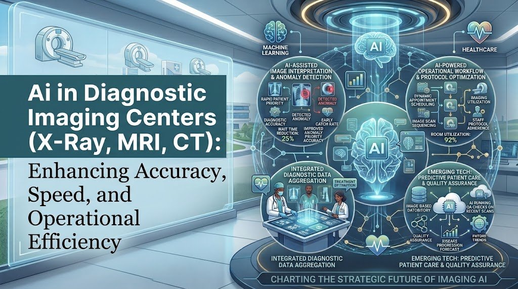
Diagnostic imaging centers sit at the intersection of healthcare, advanced hardware, digital data, and clinical decision-making. X-ray, CT, and MRI systems generate large amounts of image data that must be acquired, processed, interpreted, documented, stored, and communicated.
Traditionally, radiologists have performed most image interpretation manually, supported by PACS, reporting software, clinical information systems, and specialized visualization tools. AI is changing this workflow by adding algorithms that can identify patterns, prioritize cases, perform measurements, reconstruct images, compare studies, and prepare information for professional review.
The transformation is important because AI does not have to replace the radiologist to create value. In many practical implementations, AI acts as an additional layer between image acquisition and professional interpretation.
The system can identify potentially urgent findings and move those examinations higher in the worklist. It can automatically measure anatomical structures, highlight suspicious areas, calculate quantitative biomarkers, or prepare structured information for the radiologist.
This creates a new model for diagnostic imaging:
IMAGE ACQUISITION
X-ray, CT, MRI and related imaging data
AI PROCESSING
Detection, reconstruction, segmentation and analysis
CLINICAL WORKFLOW
Triage, measurements and reporting support
RADIOLOGIST REVIEW
Professional interpretation and final decision
Why Medical Imaging Is a Strong AI Opportunity
Medical imaging is particularly suitable for AI because the input is highly structured and many diagnostic tasks involve identifying visual patterns.
A single imaging examination can contain hundreds or thousands of individual images. A CT or MRI study may require a radiologist to review multiple anatomical regions and sequences before producing a final interpretation.
AI can help reduce repetitive analysis by identifying areas that deserve attention.
The technology can also convert images into structured measurements that would otherwise require manual work.
Common AI functions include:
- Abnormality detection.
- Image classification.
- Lesion detection.
- Organ segmentation.
- Tumor measurement.
- Fracture detection.
- Intracranial hemorrhage detection.
- Pulmonary embolism detection.
- Pneumothorax detection.
- Stroke-related triage.
- Cardiovascular imaging analysis.
- Bone-density estimation from CT.
- Body-composition measurement.
- Image reconstruction and denoising.
- Radiology report assistance.
- Follow-up recommendation tracking.
The FDA’s AI-enabled medical-device database demonstrates how large the medical-imaging AI ecosystem has become. Current FDA listings include numerous radiology products covering areas such as CT, mammography, ultrasound, image segmentation, and other imaging applications.
Source: FDA AI-Enabled Medical Devices
AI in X-Ray Imaging
X-ray is one of the most widely used imaging technologies and is an important environment for AI-assisted interpretation.
Chest X-rays, for example, can contain findings related to pneumonia, pneumothorax, pleural effusion, pulmonary edema, fractures, masses, and other conditions.
AI systems can analyze X-ray images and identify patterns that may require closer attention.
The most useful application in many imaging centers is not necessarily autonomous diagnosis. It is prioritization and decision support.
For example, if an AI system identifies a possible pneumothorax in a chest X-ray, the examination could be moved higher in the radiologist’s worklist so that it receives earlier attention.
This approach is particularly useful in high-volume imaging environments where urgent cases compete with routine examinations.
The American College of Radiology currently lists imaging AI use cases involving conditions such as pneumothorax, intracranial hemorrhage, pulmonary embolism, large-vessel occlusion, pleural effusion, and other findings.
Source: American College of Radiology Define-AI use cases
AI X-Ray Workflow
↓
AI IMAGE ANALYSIS
↓
POSSIBLE ABNORMALITY IDENTIFIED
↓
URGENT CASE PRIORITIZATION
↓
RADIOLOGIST INTERPRETATION
↓
FINAL REPORT
This workflow preserves the radiologist as the final clinical decision-maker while allowing AI to support prioritization.
AI in CT Imaging
Computed tomography creates highly detailed cross-sectional images and produces substantial amounts of data. AI can therefore provide value at multiple stages of the CT workflow.
Potential applications include detection, segmentation, reconstruction, quantification, workflow prioritization, and opportunistic screening.
AI can help identify findings such as:
- Intracranial hemorrhage.
- Pulmonary embolism.
- Large-vessel occlusion.
- Lung nodules.
- Fractures.
- Abdominal abnormalities.
- Organ lesions.
- Bone abnormalities.
- Cardiovascular calcification.
- Hepatic steatosis.
One of the major advantages of CT is that AI can also extract quantitative information from examinations that were originally performed for another reason.
For example, a CT examination can potentially be analyzed for bone mineral density, visceral fat, muscle mass, coronary artery calcification, or liver fat.
This creates an important concept known as opportunistic screening.
Instead of requiring an additional examination, AI can analyze information that already exists in a patient’s imaging data.
The ACR has identified opportunistic applications including bone mineral density, atherosclerotic calcification, and hepatic steatosis as areas where AI can extend the value of existing imaging.
Source: ACR, The Practice of AI
AI in MRI
MRI presents a different AI opportunity because MRI examinations can contain multiple sequences and require careful image acquisition.
AI can support MRI by improving image reconstruction, reducing noise, accelerating acquisition, performing segmentation, and assisting with disease detection.
One important area is accelerated MRI.
Traditional MRI can require significant acquisition time because multiple sequences must be collected. AI-based reconstruction methods can potentially reduce the amount of raw data required or reconstruct high-quality images from accelerated acquisitions.
This can create operational benefits for imaging centers.
Potential benefits include:
- Shorter scan times.
- Improved patient comfort.
- Higher scanner utilization.
- Potentially increased daily examination capacity.
- Reduced motion-related problems in selected workflows.
- Faster image reconstruction.
AI can also support segmentation of organs, tumors, cardiac structures, brain structures, and other anatomical regions.
Automated segmentation is particularly valuable when measurements need to be repeated over time.
For example, a radiologist or specialist may need to compare tumor volume across several MRI examinations. AI can assist with consistent segmentation and measurement.
AI-Powered Image Reconstruction
One of the most important developments in imaging AI is the movement from post-acquisition interpretation toward AI-assisted image formation itself.
Traditional reconstruction methods convert raw scanner data into images using established mathematical techniques. AI-based reconstruction can learn patterns that help produce clinically useful images from limited or noisy data.
This is especially important for CT and MRI.
AI reconstruction can potentially improve image quality while allowing imaging centers to explore lower-dose CT protocols or faster MRI acquisition strategies.
However, image quality cannot be evaluated only by visual appearance.
The reconstructed image must remain clinically reliable and preserve relevant diagnostic information.
This means AI reconstruction should be evaluated through both technical measurements and clinical validation.
AI for Radiology Triage
Radiology triage is one of the most practical applications of AI.
Imaging centers frequently manage examinations with very different levels of urgency. A routine follow-up scan and a potentially life-threatening intracranial hemorrhage may enter the same general workflow.
AI can help identify examinations that potentially contain urgent findings and move them higher in the worklist.
The objective is not necessarily to produce the final diagnosis.
The objective is to reduce the time between image acquisition and professional review for cases that may require urgent attention.
| AI Finding | Possible Workflow Action | Clinical Purpose |
|---|---|---|
| Intracranial hemorrhage | Prioritize CT | Earlier radiologist review |
| Pulmonary embolism | Prioritize CT | Reduce delay in review |
| Pneumothorax | Prioritize X-ray | Flag potentially urgent finding |
| Large-vessel occlusion | Urgent workflow alert | Support stroke pathway |
| Fracture | Worklist prioritization | Support timely interpretation |
What Does Clinical Research Say?
Evidence is moving beyond retrospective datasets toward clinical evaluation.
A 2026 scoping review of randomized controlled trials evaluating AI tools for radiology diagnosis found that AI systems were mainly used as clinician-facing decision aids. Across the RCT evidence mapped by the review, AI was generally associated with higher sensitivity or lesion-detection rates and shorter image-processing time, although outcomes varied between settings and applications.
This is important because high performance on an internal dataset does not automatically prove clinical usefulness.
An imaging center should ask whether the AI system improves an actual workflow outcome.
Useful questions include:
- Does it reduce time to urgent case review?
- Does it improve lesion detection?
- Does it reduce reporting workload?
- Does it improve measurement consistency?
- Does it reduce unnecessary repeat work?
- Does it improve patient throughput?
- Does it maintain performance across different scanners?
- Does it work reliably across patient populations?
The Generalizability Problem in Imaging AI
One of the biggest challenges in diagnostic imaging AI is generalizability.
An AI model may perform extremely well when tested on images that resemble its training data. Performance can change when the system encounters a different hospital, scanner manufacturer, imaging protocol, patient population, image quality, or disease prevalence.
A systematic review examining generalizability of AI models across CT, MRI, and X-ray highlighted this issue as a major barrier to clinical deployment. The review specifically examined studies with internal and external validation and focused on whether models maintained performance when moved across different clinical environments.
Source: Systematic review of AI generalizability across clinical settings
This means diagnostic imaging centers should not evaluate an AI vendor only by asking for the highest reported accuracy.
They should ask how the model performed outside the environment in which it was developed.
Important validation questions include:
- Was external validation performed?
- Were multiple hospitals involved?
- Were multiple scanner manufacturers represented?
- Were different imaging protocols included?
- Was the patient population diverse?
- Were false negatives measured?
- Were false positives measured?
- Was performance evaluated after deployment?
AI Model Drift in Diagnostic Imaging
AI performance can change after deployment.
A model may perform well when initially installed but produce different results months or years later because the imaging environment changes.
Scanner hardware may be upgraded.
Imaging protocols may change.
Patient populations may change.
The AI vendor may release a new model version.
Clinical workflows may also change.
This is why AI monitoring is becoming a major part of imaging governance.
The ACR’s 2026 imaging-AI practice parameter specifically recommends AI inventories, local acceptance testing, ongoing performance monitoring, drift monitoring, safety monitoring, and predefined stop rules.
Source: ACR first Practice Parameter for Imaging AI
AI Quality Assurance in Imaging Centers
The ACR has created ARCH-AI, a quality-assurance program designed specifically for radiology facilities using AI.
Its framework includes governance, AI inventory, testing, workflow integration, and monitoring.
ACR’s Assess-AI initiative also focuses on measuring real-world AI performance after deployment.
This represents an important shift in healthcare AI.
The question is no longer simply:
“Does this AI model work?”
The more useful question is:
“Does this AI model continue to work correctly in our imaging environment?”
The ACR describes Assess-AI as a nationwide AI quality registry designed to monitor algorithm performance in clinical practice.
Source: ACR Assess-AI and Imaging AI Practice Parameter
AI in Radiology Reporting
Generative AI is opening another major area of opportunity.
Traditional radiology reporting requires the radiologist to review images, interpret findings, compare previous examinations, organize the report, communicate important findings, and document recommendations.
Generative AI can assist with parts of this process.
Potential applications include:
- Drafting report impressions.
- Summarizing findings.
- Comparing current and previous examinations.
- Converting structured findings into report language.
- Checking report completeness.
- Generating patient-friendly explanations.
- Identifying follow-up recommendations.
- Extracting structured information from reports.
The role should remain assistive.
A generated report should be reviewed and approved by an appropriately qualified professional before becoming the final clinical record.
The ACR notes that large language models, vision-language models, and foundation models are expanding AI from narrow triage tools toward report-generation and broader workflow support. It also emphasizes that AI is probabilistic, meaning its behavior can vary and therefore requires critical assessment and monitoring.
Source: ACR, The Practice of AI
AI for Structured Reporting
AI can also help convert narrative radiology findings into structured information.
For example, an imaging center may want to track:
- Tumor measurements.
- Lung nodule size.
- Lesion location.
- Fracture characteristics.
- Organ volume.
- Bone density measurements.
- Follow-up recommendations.
Structured data can improve longitudinal analysis.
It can also support clinical research and population-level analytics.
Instead of having important information trapped inside free-text reports, AI can help convert selected findings into structured data fields.
AI and Incidental Findings
Medical imaging frequently identifies findings that were not the original reason for the examination.
These incidental findings can require follow-up.
AI can potentially identify report recommendations and help ensure that recommended follow-up does not disappear after the patient leaves the imaging center.
The ACR’s AI use-case directory includes applications focused on tracking radiology recommendations, ensuring referring clinicians read reports, and improving patient follow-up for findings such as pulmonary nodules.
Source: ACR Define-AI use cases
This creates an important distinction.
AI does not only have to interpret images.
It can also help close the loop after interpretation.
AI for Patient Communication
Imaging centers can use generative AI to improve communication before and after examinations.
Potential applications include:
- Explaining preparation requirements.
- Answering common procedure questions.
- Providing multilingual information.
- Explaining general imaging terminology.
- Communicating appointment instructions.
- Updating patients about scheduling delays.
- Explaining general follow-up instructions already approved by clinicians.
The ACR’s AI use-case directory includes patient-facing applications such as chatbots for radiology procedure questions, translation of radiology reports into lay language, patient follow-up, and communication regarding incidental findings.
Source: ACR Define-AI patient-facing use cases
AI for Scheduling and Imaging Center Operations
AI can also improve the operational side of diagnostic imaging.
Imaging centers must coordinate scanners, technologists, radiologists, appointment slots, contrast requirements, patient preparation, equipment availability, and emergency examinations.
Machine-learning systems can analyze historical scheduling patterns to predict demand.
Potential applications include:
- Predicting appointment no-shows.
- Forecasting daily imaging volume.
- Optimizing scanner utilization.
- Predicting staffing requirements.
- Identifying scheduling bottlenecks.
- Optimizing appointment sequencing.
- Predicting equipment downtime.
- Improving patient flow.
The ACR’s imaging AI use-case directory explicitly includes applications for predicting radiology appointment no-shows and forecasting imaging volume for staffing optimization.
Source: ACR Define-AI operational use cases
AI for Radiology Worklist Optimization
A high-volume imaging center can have hundreds of examinations waiting for interpretation.
Not every examination has the same urgency or complexity.
AI can help organize worklists using multiple signals.
Clinical Priority
Potentially urgent findings can be flagged for earlier review.
Study Complexity
AI can help identify examinations requiring additional attention.
Workflow Load
Work can be distributed according to availability and workload.
Follow-Up
Prior examinations and recommendations can be incorporated into workflow.
The objective should be to improve the overall system rather than simply make radiologists read more examinations.
AI in PACS and RIS Integration
AI creates the greatest value when it is integrated into existing imaging workflows.
A disconnected AI application that requires radiologists to manually upload images, wait for results, and then return to PACS can create additional friction.
A better architecture integrates AI with existing systems.
MODALITY
X-RAY / CT / MRI
↓
PACS / DICOM
↓
AI ORCHESTRATION LAYER
↓
DETECTION + SEGMENTATION + RECONSTRUCTION + QUANTIFICATION
↓
AI RESULTS
↓
RADIOLOGIST WORKSTATION
↓
REPORTING / RIS / EHR
The integration layer should support appropriate standards and secure data exchange.
It should also preserve auditability so the organization can determine which AI system generated a result, which model version was used, when it ran, and whether a professional reviewed the output.
AI Capability Map for Diagnostic Imaging Centers
| AI Capability | X-Ray | CT | MRI | Business Value |
|---|---|---|---|---|
| Abnormality detection | High | High | High | Decision support |
| Urgent triage | High | High | Moderate | Faster review |
| Segmentation | Moderate | High | High | Quantification |
| Image reconstruction | Low | High | High | Quality and efficiency |
| Report assistance | High | High | High | Productivity |
| Opportunistic screening | Moderate | High | High | Additional insights |
| Workflow forecasting | High | High | High | Operations |
AI and Radiology Productivity
AI can improve productivity in several ways, but productivity should not simply mean increasing the number of studies interpreted by every radiologist.
A better objective is to reduce unnecessary cognitive and administrative workload.
Examples include:
- Automatically prioritizing urgent examinations.
- Reducing repetitive measurements.
- Automating routine segmentation.
- Preparing structured findings.
- Comparing prior examinations.
- Preparing report drafts.
- Identifying missing report elements.
- Tracking recommendations.
- Reducing repetitive data entry.
This distinction matters because radiology is a high-responsibility environment.
An AI system that simply increases reading speed without maintaining diagnostic quality may create new risks.
AI and Diagnostic Accuracy
Diagnostic accuracy is one of the most attractive promises of medical imaging AI, but it should be evaluated carefully.
A model can have excellent sensitivity while generating too many false positives.
Another model can have high specificity but miss clinically important abnormalities.
Therefore, imaging centers should evaluate multiple performance metrics.
| Metric | Why It Matters |
|---|---|
| Sensitivity | How well the system identifies relevant positive cases. |
| Specificity | How well the system avoids incorrectly flagging negative cases. |
| PPV | How often positive AI results represent true positives in the evaluated population. |
| NPV | How often negative AI results represent true negatives. |
| AUC | Overall discrimination across thresholds. |
| Turnaround Time | Measures workflow impact rather than only technical accuracy. |
The correct metrics depend on the intended clinical use.
Bias and Equity in Imaging AI
Imaging AI can inherit biases from its training data.
If a dataset does not adequately represent certain populations, scanner types, age groups, or clinical environments, model performance may differ after deployment.
A responsible AI program should therefore evaluate performance across relevant subgroups.
This is particularly important for large diagnostic imaging networks operating across different hospitals and geographic regions.
A recent systematic review of responsible AI in medical imaging emphasized that safe AI requires more than high diagnostic accuracy. The review identified transparency, subgroup performance, privacy, uncertainty calibration, and clinical trustworthiness as important components of responsible imaging AI.
Source: Responsible artificial intelligence in medical imaging systematic review
AI Security and Patient Privacy
Diagnostic images are highly sensitive medical data.
An imaging AI architecture should therefore protect both the image and associated metadata.
Security controls should include:
- Role-based access control.
- Encryption during transmission.
- Encryption at rest where appropriate.
- Audit logs.
- Secure API authentication.
- Controlled vendor access.
- Data-retention policies.
- De-identification for research datasets.
- Model-access monitoring.
- Incident-response procedures.
The ACR’s AI practice parameter specifically emphasizes patient privacy, access controls, logging, governance, and continuous monitoring as part of responsible AI implementation.
Source: ACR Imaging AI Practice Parameter
FDA Regulatory Considerations
AI used for medical imaging may fall within medical-device regulation depending on its intended use and functionality.
The FDA maintains an AI-enabled medical-device list and explains that listed devices have met applicable premarket requirements, including evaluation of safety and effectiveness appropriate to their intended use and technological characteristics.
Source: FDA AI-Enabled Medical Devices
The regulatory environment is also evolving as AI systems become more adaptive and generative.
The FDA’s 2026 discussion paper on generative-AI-enabled medical devices highlights that generative systems can introduce risks that differ from traditional AI-enabled medical devices.
Source: FDA discussion paper on generative AI-enabled medical devices
Imaging centers should therefore evaluate the regulatory status of an AI product according to its actual intended use rather than assuming that every AI feature has the same regulatory requirements.
AI Governance Framework for Diagnostic Imaging Centers
A mature imaging center should create an AI governance structure before deploying multiple clinical AI systems.
Governance
Clinical, technical, compliance and administrative ownership.
Validation
Local testing before clinical deployment.
Monitoring
Continuous measurement of performance and drift.
Safety
Defined escalation procedures and stop rules.
The ACR’s 2026 practice parameter recommends an AI governance group, inventory of AI tools and versions, local acceptance testing, real-world monitoring, privacy controls, and defined stop rules.
Source: ACR Practice Parameter for Imaging AI
AI Maturity Model for Imaging Centers
| Stage | AI Capability | Typical Examples |
|---|---|---|
| Foundation | Digital workflow | PACS, RIS, digital reporting |
| Assisted | Narrow AI | Triage and abnormality detection |
| Integrated | AI workflow integration | PACS-integrated AI and automated measurements |
| Intelligent | Predictive and generative AI | Report assistance, forecasting, patient communication |
| AI-Native | Closed-loop intelligence | Continuous monitoring, personalized workflows, multimodal AI |
High-Value AI Use Cases
The highest-value opportunities generally combine measurable clinical or operational benefits with manageable implementation complexity.
- Urgent case triage.
- Abnormality detection.
- Automated measurements.
- Organ and lesion segmentation.
- AI-assisted reporting.
- Prior-study comparison.
- Imaging recommendation follow-up.
- Scheduling optimization.
- No-show prediction.
- Scanner utilization forecasting.
- AI-assisted image reconstruction.
- Opportunistic screening from existing CT examinations.
These applications can be introduced independently and later connected into a broader imaging-intelligence platform.
Higher-Risk AI Use Cases
Some applications require much stronger validation and governance because errors can directly affect clinical decisions.
These include:
- Autonomous diagnosis.
- AI-generated final reports without professional review.
- AI-generated treatment recommendations.
- AI-based clinical decisions without escalation mechanisms.
- Automated dismissal of negative examinations without human oversight.
- AI systems that continuously change their behavior without monitoring.
The higher the clinical consequence of an incorrect result, the stronger the validation and oversight requirements should be.
AI Implementation Roadmap
Define the Problem
The imaging center should first identify a measurable problem such as delayed urgent-case review, excessive reporting workload, poor follow-up tracking, long MRI acquisition times, or underutilized scanner capacity.
Evaluate the Evidence
The organization should examine peer-reviewed evidence, regulatory status, external validation, local compatibility, and real-world performance.
Run Local Acceptance Testing
An AI product should be evaluated against the imaging center’s own scanners, protocols, patient population, and workflow before broad deployment.
Integrate with PACS and RIS
AI should appear within the existing workflow whenever possible rather than creating another disconnected system.
Train the Workforce
Radiologists, technologists, administrators, IT teams, and other relevant professionals should understand what the system does, what it does not do, and how AI results should be handled.
Monitor Continuously
The center should track performance after deployment and establish procedures for model drift, vendor updates, unexpected behavior, and safety incidents.
Future of AI in X-Ray, CT and MRI
Multimodal Imaging AI
Future systems will increasingly combine imaging with clinical history, laboratory information, prior reports, demographics, and other data.
Instead of asking only what appears in an image, multimodal systems can potentially interpret the image within the patient’s broader clinical context.
Vision-Language Models
Vision-language models can connect medical images with natural-language information.
This could support report generation, image-question answering, structured findings, patient-friendly explanations, and research data extraction.
However, the probabilistic nature of generative systems makes professional review and monitoring particularly important.
AI-Native Radiology Workstations
The future workstation may no longer be a passive image viewer.
It could actively organize cases, display AI findings, compare prior examinations, present quantitative measurements, identify follow-up recommendations, and prepare a report draft.
The radiologist would remain responsible for interpretation while AI handles more of the repetitive information-processing work.
Continuous AI Quality Monitoring
As imaging centers deploy multiple AI systems, monitoring platforms will become increasingly important.
ACR’s Assess-AI initiative represents an early example of this direction, focusing on real-world performance monitoring rather than relying only on pre-deployment validation.
Opportunistic Screening Will Expand
CT and MRI examinations contain information beyond the original clinical question.
AI can potentially extract additional biomarkers from existing scans, creating new opportunities for cardiovascular risk assessment, metabolic assessment, bone health, body composition, and other areas.
AI Will Become Part of the Imaging Infrastructure
The most mature imaging centers will not treat AI as a collection of separate tools.
They will build an AI infrastructure layer that manages multiple models, routes examinations, monitors outputs, records model versions, tracks performance, and connects results with clinical systems.
Recommended AI Architecture
Imaging Layer
X-ray, CT, MRI, DICOM, scanner metadata and acquisition information.
Integration Layer
PACS, RIS, EHR, DICOM routing, APIs and workflow orchestration.
AI Layer
Detection, segmentation, reconstruction, prediction, NLP and multimodal models.
Clinical Layer
Radiologist workstation, reporting, alerts, measurements and follow-up workflows.
Governance Layer
Monitoring, audit logs, model inventory, validation, privacy and safety controls.
AI Opportunity Matrix
| Use Case | Technology | Potential Impact | Risk Level |
|---|---|---|---|
| Urgent triage | Computer vision | Faster review | Moderate |
| Segmentation | Deep learning | Measurement automation | Moderate |
| Image reconstruction | Deep learning | Speed and image quality | Moderate to high |
| Report drafting | LLM/VLM | Productivity | High |
| Opportunistic screening | Computer vision + ML | Additional clinical insights | Moderate to high |
| Autonomous diagnosis | Advanced AI | Potential automation | Very high |
What Healthcare Startups Can Build
Diagnostic imaging is an attractive area for healthcare AI startups because there are many narrowly defined problems with measurable outcomes.
The strongest startup opportunities are likely to be specialized rather than generic.
Potential products include:
- AI triage platforms for emergency imaging.
- AI-assisted CT lung analysis.
- Automated MRI segmentation platforms.
- AI image reconstruction solutions.
- Radiology reporting copilots.
- Automated prior-study comparison.
- Incidental-finding follow-up systems.
- AI quality-monitoring platforms.
- Radiology workflow orchestration platforms.
- Opportunistic screening systems.
- AI-powered imaging center scheduling.
- AI no-show prediction.
- Multimodal radiology assistants.
- Patient-friendly radiology communication systems.
The strongest products should solve a clearly defined workflow problem and demonstrate measurable value.
Legacy Imaging Center Modernization
Many imaging centers have modern scanners but fragmented software environments.
The modernization opportunity is therefore not always to replace the scanner or PACS.
AI can act as a modernization layer around existing systems.
A legacy imaging center could gradually introduce:
- Automated worklist prioritization.
- AI detection tools.
- Automated measurements.
- Reporting assistance.
- Patient communication.
- Follow-up tracking.
- Scheduling intelligence.
- AI governance and monitoring.
This incremental approach can reduce disruption and allow organizations to measure ROI at each stage.
How to Measure AI ROI
AI investment should be evaluated using more than software cost.
An imaging center should consider:
| ROI Area | Example KPI |
|---|---|
| Clinical | Detection performance, turnaround time, follow-up completion |
| Radiologist | Reporting time, workload, repetitive-task reduction |
| Patient | Waiting time, appointment availability, communication quality |
| Operations | Scanner utilization, no-show rate, throughput |
| Financial | Cost per examination, capacity, revenue and AI operating cost |
A successful AI project should produce measurable improvement in one or more of these areas without compromising clinical quality.
Frequently Asked Questions
How is AI used in diagnostic imaging centers?
AI can analyze X-ray, CT, and MRI images for abnormalities, prioritize urgent cases, perform segmentation and measurements, improve image reconstruction, support radiology reporting, predict operational demand, track follow-up recommendations, and assist with patient communication.
Can AI diagnose X-rays?
AI systems can detect and classify specific abnormalities in X-ray images, but the exact role depends on the intended use, validation, and regulatory status of the system. Many practical applications are designed as decision-support or triage tools rather than complete replacement for professional interpretation.
How does AI help CT scans?
AI can support CT interpretation, urgent triage, lesion detection, segmentation, quantitative analysis, image reconstruction, and opportunistic screening for additional findings.
How does AI help MRI?
AI can assist with MRI reconstruction, image denoising, acquisition acceleration, segmentation, quantitative analysis, lesion detection, and reporting support.
Can AI reduce MRI scan time?
AI-based reconstruction and acquisition techniques can potentially support faster MRI workflows, although the actual benefit depends on the scanner, sequence, AI technology, protocol, and clinical requirements.
Can AI improve radiology reporting?
Yes. Generative AI and other language-based technologies can assist with report drafting, summarization, structured findings, prior-study comparison, and patient-friendly explanations. Final clinical reports should remain subject to appropriate professional review.
What is AI triage in radiology?
AI triage analyzes imaging examinations and identifies cases that may contain urgent findings. These cases can then be prioritized in the radiologist’s worklist so they receive earlier professional review.
What is opportunistic screening in medical imaging?
Opportunistic screening uses imaging that was already acquired for one clinical purpose to identify additional measurable findings. CT is particularly promising because a single examination contains information about multiple anatomical and physiological characteristics.
What is the biggest challenge with medical imaging AI?
One of the biggest challenges is generalizability. A model that performs well in one hospital or dataset may perform differently on images from other hospitals, scanners, protocols, or patient populations.
How should imaging centers monitor AI?
They should maintain an AI inventory, perform local acceptance testing, monitor real-world performance, track model versions, evaluate drift, maintain appropriate privacy controls, and establish escalation or stop rules.
Can AI replace radiologists?
Current practical evidence and professional guidance support AI as an assistive technology rather than assuming complete replacement of radiologists. AI is increasingly integrated into radiology workflows for triage, detection, measurement, reconstruction, reporting, and decision support, while professional interpretation remains important.
Research Sources
- American College of Radiology, First Practice Parameter for Imaging AI: ACR’s 2026 framework covering AI selection, governance, local testing, monitoring, privacy, performance drift, and quality improvement. Original source
- American College of Radiology, The Practice of AI: 2026 overview of radiology AI adoption, including triage, segmentation, opportunistic screening, LLMs, vision-language models, and AI monitoring. Original source
- ACR Define-AI Use Cases: Directory of clinical, operational, and patient-facing AI applications in medical imaging. Original source
- ACR AI in Your Practice: Resources covering ARCH-AI, AI Central, Assess-AI, datasets, use cases, and imaging AI implementation. Original source
- FDA AI-Enabled Medical Devices: FDA database and regulatory information for AI-enabled medical devices authorized for marketing in the United States. Original source
- FDA AI/ML Medical Device Research Program: FDA research covering image acquisition, image processing, early disease detection, diagnosis, prognosis, risk assessment, and personalized diagnostics. Original source
- FDA Digital Health Guidance: Current FDA guidance covering AI-enabled device software, clinical decision support, cybersecurity, and lifecycle considerations. Original source
- FDA Generative AI Medical Device Discussion Paper: FDA discussion of regulatory considerations and risks associated with generative-AI-enabled medical devices. Original source
- JAMA Network Open, FDA Approval of AI and ML Devices in Radiology: Systematic review of FDA-cleared AI/ML devices in radiology and their testing landscape. Original source
- Randomized Controlled Trial Evidence for AI in Radiological Diagnosis: 2026 scoping review of RCT evidence evaluating AI tools for imaging-based diagnosis. Original source
- Generalizability of AI in Radiology: Systematic review examining AI performance across different clinical settings, including CT, MRI, and X-ray. Original source
- Responsible AI in Medical Imaging: Systematic review addressing transparency, subgroup performance, privacy, uncertainty, and clinical trustworthiness in imaging AI. Original source
Final Perspective
AI is moving diagnostic imaging from a primarily image-viewing workflow toward a more intelligent, data-driven environment.
X-ray, CT, and MRI centers can use AI at almost every stage of the imaging journey, from acquisition and reconstruction to detection, triage, measurement, reporting, follow-up, and operational planning.
The strongest near-term opportunities are not necessarily fully autonomous diagnosis. They are targeted systems that solve measurable problems.
An AI model that moves a potentially critical CT examination to the top of a worklist can create value.
An AI system that automatically measures a lesion across multiple examinations can create value.
An AI reconstruction system that supports faster MRI acquisition can create value.
A reporting assistant that reduces repetitive documentation can create value.
A follow-up system that prevents an important imaging recommendation from being lost can create value.
The next stage will be the integration of these capabilities into a common imaging intelligence layer.

Leave a Reply