
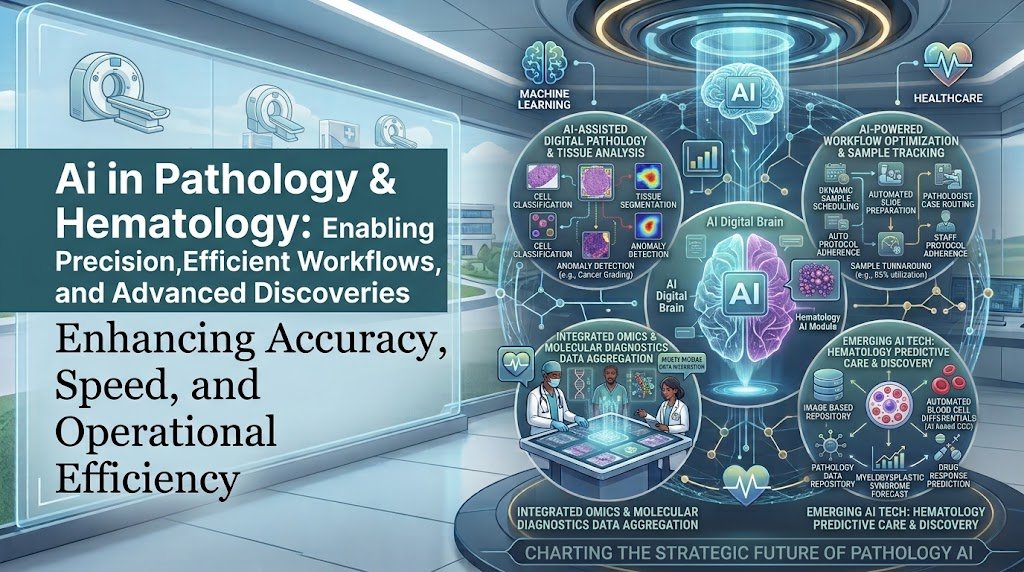
Primary topic: AI in Pathology & Hematology
Research focus: Digital pathology, whole-slide imaging, computational pathology, pathology foundation models, blood-smear analysis, leukemia detection, bone-marrow analysis, flow cytometry, molecular hematopathology, multimodal AI, laboratory automation, clinical validation, governance, and healthcare AI implementation.
Why AI in Pathology and Hematology Matters Now
Pathology and hematology are unusually rich sources of structured and unstructured clinical data. A single cancer case can involve tissue morphology, whole-slide images, immunohistochemistry, laboratory values, blood-smear findings, flow cytometry, cytogenetics, molecular testing, previous reports, and clinical history. The diagnostic challenge is therefore not simply recognizing an image. It is integrating many different forms of evidence correctly.
AI is particularly well suited to this environment because modern machine-learning systems can recognize patterns in large image datasets, process high-dimensional laboratory information, quantify features that are difficult to measure manually, and combine information from multiple sources. Recent reviews describe applications across morphology, flow cytometry, genetics, haemostasis, diagnosis, risk stratification, and treatment-response prediction.
Source: Artificial Intelligence in Haematologic Diagnostics: Current Applications and Future Perspectives
Whole-slide images, tissue segmentation, tumor detection, biomarker quantification, cancer classification and prognosis.
CBC analysis, peripheral blood smears, bone marrow, cell morphology, flow cytometry and treatment monitoring.
Cytogenetics, sequencing, mutations, biomarkers and genotype-phenotype relationships.
Computer vision, machine learning, foundation models, multimodal AI and workflow automation.
Digital Pathology Is the Foundation of Modern Pathology AI
Traditional pathology relies on glass slides and microscopes. Digital pathology converts those slides into high-resolution digital files that can be stored, transferred, searched, measured and analyzed computationally. Once tissue becomes machine-readable, AI can operate on it at a scale that would be difficult to achieve through manual microscopy alone.
The clinical importance of this transition is significant because digital pathology creates the infrastructure required for computational pathology. AI cannot analyze a physical glass slide directly. It needs a standardized digital representation, reliable image quality, appropriate metadata, and an environment in which results can be reviewed by a qualified specialist.
A 2024 systematic review and meta-analysis evaluated AI-based diagnostic accuracy in digital pathology. The review included 100 diagnostic accuracy studies, with 48 studies contributing to quantitative meta-analysis and more than 152,000 whole-slide images. The pooled research reported high diagnostic performance, but the authors also identified important methodological concerns that limit direct translation into routine clinical practice.
96.3%
93.3%
99%
The most important lesson is that high accuracy on paper does not automatically mean clinical readiness. A model can perform well on images that resemble its training data and then lose performance when scanner characteristics, staining methods, patient populations, disease prevalence or laboratory workflows change.
Whole-Slide Imaging Creates a Unique AI Challenge
Whole-slide images are enormous compared with ordinary medical images. A single slide can contain millions or billions of pixels, which makes direct end-to-end processing computationally expensive. For this reason, many pathology AI systems divide slides into smaller image tiles, extract features from those tiles, and then combine the information to make a slide-level prediction.
This approach is often called multiple-instance learning. Instead of requiring a pathologist to manually label every microscopic region, the model can learn relationships between groups of image regions and a final slide-level diagnosis. This is one reason foundation models and advanced attention mechanisms are becoming increasingly important in computational pathology.
Source: Aligning knowledge concepts to whole slide images for precise histopathology image analysis
Recent computational pathology reviews describe the field as moving from basic digitization toward segmentation, tumor classification, prognosis prediction, biomarker discovery, foundation models and generative AI. The same research identifies computational scaling, noisy annotations, interpretability, data security, reliability, reproducibility and domain shift as major barriers.
Pathology Foundation Models Are Changing the AI Architecture
Earlier pathology AI systems were usually built for one narrow task. One model might detect prostate cancer, another might classify breast cancer, and another might quantify a biomarker. This approach can work well, but it creates a fragmented AI environment with many separate models that require separate training, validation and maintenance.
Foundation models attempt to solve part of this problem by learning broad visual representations from very large pathology datasets. The resulting representation can then be adapted to different downstream tasks.
CHIEF is one of the clearest examples. Researchers developed the model using 60,530 whole-slide images covering 19 anatomical sites and approximately 44 terabytes of high-resolution pathology data. The model was subsequently evaluated using 19,491 whole-slide images from 32 independent slide sets across 24 hospitals and cohorts.
The study reported that CHIEF could support cancer-cell detection, tumor-origin identification, molecular-profile characterization and prognostic prediction. It also reported improvements over existing deep-learning methods of up to 36.1% in evaluated tasks.
Source: A pathology foundation model for cancer diagnosis and prognosis prediction
Whole-slide images used to develop CHIEF.
Anatomical sites represented in the training data.
High-resolution pathology imaging data used during pretraining.
Hospitals and cohorts represented in independent validation.
Virchow is another major pathology foundation model. The research demonstrated that a large general-purpose pathology model could be used for pan-cancer detection and could also help with rare cancer variants when less labeled training data were available.
Source: A foundation model for clinical-grade computational pathology and rare cancers detection
These developments matter for healthcare startups because the future product may not be a single-purpose “cancer detector.” A more valuable product could provide a reusable pathology intelligence layer that supports multiple diseases, biomarkers and workflows while preserving strict clinical controls.
What Recent Research Says About Foundation Models in Clinical Pathology
The gap between research performance and real-world deployment is still substantial. A 2026 systematic review of foundation models and AI agents in digital pathology identified 42 eligible studies. Foundation models showed AUC values of approximately 0.85–0.97 across downstream diagnostic tasks, but only six studies, or 14.3%, conducted multicenter validation. Prospective studies accounted for less than 5% of the reviewed evidence.
The review also found that AI agents were generally operating at early levels of workflow integration and identified four major implementation barriers: data heterogeneity, limited explainability, insufficient workflow-level validation and regulatory gaps.
Foundation-model / AI-agent studies reviewed.
Studies with multicenter validation.
Prospective studies.
Data heterogeneity, explainability, workflow validation and regulation.
AI for Tissue Detection and Tumor Classification
One of the most mature pathology applications is identifying abnormal tissue and tumor regions. AI can scan large areas of a whole-slide image and highlight regions that deserve closer inspection. This can reduce the amount of visual searching required from pathologists and can create quantitative measurements that are more reproducible than purely subjective visual estimates.
- Tumor versus non-tumor tissue classification.
- Detection of suspicious regions.
- Nuclear and cellular segmentation.
- Tissue-component classification.
- Mitotic-figure detection.
- Tumor burden estimation.
- Cell-density measurement.
- Biomarker quantification.
- Histologic pattern recognition.
- Case-level triage.
The important development is that AI can transform pathology from qualitative description toward quantitative measurement. Instead of simply stating that a tissue feature is “high” or “low,” computational systems can calculate measurements across thousands or millions of cells.
AI-Based Biomarker Quantification
Biomarker assessment is another strong area because many biomarkers are evaluated by estimating staining intensity, percentage of positive cells or spatial distribution. Manual scoring can be affected by interobserver variation and can be time-consuming in high-volume environments.
AI can standardize the measurement process by detecting cells, classifying staining patterns, quantifying positive and negative populations, and producing reproducible numerical outputs for pathologist review.
Foundation models are also increasingly being investigated for biomarker-expression prediction directly from histopathology images. CHIEF, for example, demonstrated the ability to predict molecular characteristics from pathology images in multiple cancer contexts.
Source: CHIEF pathology foundation model research
AI in Hematology Is More Than Blood-Smear Recognition
Hematology is a multimodal discipline. Blood counts, cell morphology, flow cytometry, cytogenetics, sequencing, coagulation tests and clinical history can all contribute to a diagnosis. This makes hematology an especially strong environment for multimodal AI.
Recent reviews describe AI applications across peripheral blood and bone-marrow cytomorphology, flow cytometry, genetics, haemostasis and automated interpretation. They also emphasize that final interpretation still requires expert hematologist involvement because current systems face validation, compatibility, bias and regulatory limitations.
Source: Artificial Intelligence in Haematologic Diagnostics: Current Applications and Future Perspectives
CBC + Blood Smear + Bone Marrow + Flow Cytometry + Cytogenetics + Molecular Data + Clinical History
↓
AI Evidence Integration Layer
↓
Risk Signals + Pattern Detection + Differential Support + Evidence Summary
↓
Hematologist / Hematopathologist Review
AI for Peripheral Blood-Smear Analysis
Peripheral blood-smear analysis is one of the clearest applications for computer vision in hematology. AI can detect and classify cells, identify unusual morphology, estimate cell populations and flag cases that require specialist review.
The practical benefit is not limited to speed. Digital morphology can also create standardized quantitative outputs and reduce some of the subjectivity associated with manual cell classification.
A review of digital pathology and AI in hematopathology identified automated peripheral-blood analysis, bone-marrow analysis, flow cytometry and hematolymphoid disease classification as important areas of development. The authors specifically described automated systems as a way to streamline workflows and potentially improve turnaround time.
Source: Digital pathology and artificial intelligence as the next chapter in diagnostic hematopathology
AI for Leukemia Detection
Leukemia detection is one of the most heavily researched AI applications in hematology. Peripheral blood-smear images contain morphological information that can be analyzed using computer vision and deep-learning models.
A systematic review and meta-analysis published on AI detection of acute myeloid leukemia from microscopic blood images found high overall accuracy and sensitivity across the analyzed studies, while also reporting substantial variation between studies. The authors concluded that future research needs more standardized reporting and performance assessment.
A separate 2025 systematic review and meta-analysis evaluated AI-based leukemia detection and classification from peripheral blood-smear images. The research highlights the growing potential of AI for automated leukemia screening and subtype classification while also showing why standardized datasets and external validation are necessary.
Source: Towards Diagnostic Intelligent Systems in Leukemia Detection and Classification
- Digital blood-smear acquisition.
- Image-quality assessment.
- Cell detection and segmentation.
- White-cell classification.
- Abnormal-cell and blast detection.
- Case-level risk prioritization.
- Integration with CBC and other laboratory values.
- Specialist review and confirmation.
AI for Acute Myeloid Leukemia and Other Myeloid Disorders
AML and myelodysplastic syndromes create a particularly interesting AI problem because diagnosis and classification depend on multiple evidence streams. AI research now covers blood and bone-marrow images, flow cytometry, molecular information, prognosis and treatment prediction.
A 2025 review of AI in myeloid malignancies described applications spanning diagnosis, prognostication and treatment prediction. Deep learning has been applied to bone-marrow smears, peripheral blood films and flow cytometry, with many studies reporting high diagnostic performance.
Another 2025 review of AML diagnostic research grouped AI approaches into three major data modalities: blood-smear image analysis, flow-cytometry interpretation and genetic-data modeling. This is important because future AML systems are likely to become multimodal instead of relying on morphology alone.
Source: Research advances in the adjunctive diagnosis of acute myeloid leukemia
AI for Bone-Marrow Pathology
Bone-marrow pathology is difficult because diagnosis requires assessment of cellularity, lineages, megakaryocytes, fibrosis, abnormal cells and spatial relationships. AI can convert these qualitative observations into quantitative measurements.
Research has explored automated characterization of cell lineages, cell counting, stromal structures and disease-associated morphology. Bone-marrow AI may also identify subtle phenotypic patterns that are difficult to quantify consistently through manual review.
A 2025 study developed an AI-based quantitative bone-marrow pathology platform for myeloproliferative neoplasms. The study included 342 total cases and analyzed features such as marrow cellularity, myeloid-to-erythroid ratio, megakaryocyte morphology and distribution, and fibrosis grading. The reported accuracy for several bone-marrow metrics was approximately 0.9, while segmentation and identification tasks achieved intersection-over-union values around 0.8.
Identify and quantify hematopoietic cell populations.
Measure morphology, density and spatial distribution.
Support reproducible grading and quantitative assessment.
Identify patterns associated with myeloproliferative disorders.
AI for Flow Cytometry
Flow cytometry creates high-dimensional data by measuring multiple markers across large populations of cells. Traditional analysis frequently relies on expert-defined gates and interpretation. AI can help automate clustering, identify abnormal populations, reduce subjective variation and support classification.
Recent hematology reviews report that AI can support flow-cytometry analysis and may reduce variability between human interpretations. Research is also moving toward integrating flow-cytometry information with morphology and molecular data.
Source: Artificial Intelligence in Haematologic Diagnostics
A 2026 review of molecular pathology and new technologies in hematologic diagnostics reported that deep learning applied to multiparameter flow cytometry has achieved performance comparable with expert review in distinguishing mature B-cell neoplasms and acute leukemias in the studies examined.
Source: Molecular Pathology, Artificial Intelligence, and New Technologies in Hematologic Diagnostics
AI for Cytogenetics and Molecular Hematopathology
Hematologic diagnosis increasingly depends on genomic and molecular information. Machine learning can assist with classification, mutation prediction, sequencing analysis, karyotype interpretation and integration of genotype with morphology.
The longer-term opportunity is not simply using AI to interpret sequencing. It is connecting molecular findings with the visual phenotype observed in blood or bone marrow. A system could potentially recognize a morphology pattern, retrieve associated molecular evidence, and present the relationship to a hematopathologist.
Recent reviews describe AI applications across genetics, high-throughput sequencing, cytogenetics and molecular hematopathology, while also highlighting the need for cross-platform validation and standardized data.
Source: Artificial Intelligence in Haematologic Diagnostics
AI for Hemostasis and Coagulation
AI opportunities extend beyond cancer and morphology. Hematology laboratories also manage coagulation and haemostasis testing, where AI can support quality control, personalized reference ranges and interpretation of complex result patterns.
These applications are less visible than cancer detection but can be operationally valuable because laboratory systems generate large volumes of structured data. AI can identify unusual patterns, detect quality-control abnormalities and support laboratory specialists in interpreting results.
Source: Artificial Intelligence in Haematologic Diagnostics: Current Applications and Future Perspectives
AI for Pathology Triage and Worklist Optimization
One of the most practical AI applications is deciding which cases should receive attention first. Instead of treating every case as equally urgent, AI can use predefined rules and validated models to identify cases that may require earlier review.
- Flag potentially malignant cases.
- Prioritize abnormal blood smears.
- Identify suspected leukemia cases.
- Route specialized cases to appropriate experts.
- Identify slides requiring additional review.
- Detect poor-quality digital slides.
- Prioritize cases approaching turnaround-time limits.
This is an important implementation strategy because triage AI can create value without making the final diagnosis. It can therefore be introduced as a decision-support layer while maintaining a strong human-in-the-loop structure.
AI for Digital Pathology Quality Control
AI can also be used before diagnostic interpretation begins. Poor focus, tissue folds, staining artifacts, scanning errors and other image-quality problems can interfere with digital pathology. Automatically detecting these problems can prevent pathologists from wasting time reviewing images that are unsuitable for reliable interpretation.
The FDA has already recognized an AI-based digital pathology image-artifact detection software device category. Its intended function is to identify specific artifacts in scanned whole-slide images so that images can undergo further evaluation before diagnostic review.
Source: FDA Product Classification: Digital Pathology Image Artifact Detection Software
This is strategically important because quality-control AI can become an early and comparatively lower-risk entry point for laboratories beginning their AI journey.
AI-Assisted Pathology Reporting
Generative AI introduces a new layer beyond image classification. A pathology copilot could summarize verified findings, retrieve relevant historical information, organize molecular results, prepare structured report sections and identify missing information for the pathologist.
However, generative AI must be tightly constrained in clinical reporting. The system should not invent microscopic findings, fabricate laboratory values or silently transform uncertainty into certainty.
Verified source data
AI evidence retrieval
AI summary
Structured draft
Pathologist verification
Multimodal AI Is the Bigger Long-Term Opportunity
Hematopathology is naturally multimodal because a final diagnosis may depend on morphology, immunophenotype, genetics and clinical context. A system that analyzes only one modality is therefore limited by design.
Modern research increasingly points toward AI systems that combine image and text information. Pathology vision-language models are being developed to connect microscopic images with natural-language descriptions, questions and clinical concepts.
PathChat is one example of this direction. The multimodal pathology copilot was trained using more than one million pathology image-caption pairs along with a large collection of curated instructions, allowing it to answer pathology-related questions using both visual and textual information.
Source: A multimodal AI system for pathology
The future architecture could therefore look less like a collection of separate AI tools and more like a central diagnostic intelligence layer.
Whole-Slide Images + Blood Smears + Bone Marrow + Flow Cytometry + CBC + Molecular Data + Clinical Context
↓
Data Normalization & Interoperability
↓
Vision Models + Tabular ML + Language Models + Foundation Models
↓
Evidence Fusion & Risk Stratification
↓
Specialist Copilot
↓
Pathologist / Hematologist Final Decision
Why Multimodal AI Is Difficult
Combining multiple datasets creates more value but also more failure points. Images may come from different scanners, laboratory data may use different conventions, molecular reports may contain different formats, and clinical records may contain incomplete or inconsistent information.
| Problem | Example | AI consequence |
|---|---|---|
| Data heterogeneity | Different scanners and laboratory systems | Performance variation |
| Missing data | No molecular result available | Incomplete evidence |
| Label noise | Disagreement between historical diagnoses | Training instability |
| Domain shift | Different population or protocol | Reduced generalizability |
| Temporal drift | New instruments or diagnostic criteria | Model degradation |
Clinical Validation Is More Important Than Benchmark Accuracy
A pathology AI system can achieve excellent performance in a research paper and still fail to deliver clinical value. The model must be validated in the laboratory where it will actually operate.
The College of American Pathologists states that image-analysis and AI systems must undergo laboratory validation before clinical use, even when the system has already received FDA approval. CAP guidance also emphasizes that validation should reflect the intended clinical setting, specimen types and diagnostic complexity.
Source: College of American Pathologists: How to Validate AI Algorithms in Anatomic Pathology
The CAP educational guidance applies whole-slide imaging validation principles to AI image-analysis systems and describes a minimum sample set of 60 cases for one application as a useful validation framework, while emphasizing that the medical director determines appropriate acceptance criteria for the intended use.
Source: CAP AI Algorithm Validation Guidance
FDA Research Shows Why Interoperability and Generalizability Matter
The FDA’s Digital Pathology Program identifies several regulatory-science gaps that directly affect AI deployment. These include the lack of standardized methods connecting technical image performance with clinical performance, interoperability challenges, image-quality assessment, reproducibility and methods for measuring AI generalizability.
Source: FDA Digital Pathology Program
This means a serious pathology AI implementation needs to evaluate more than the neural network. The scanner, image pipeline, software environment, display, integration layer, user workflow and clinical population all form part of the operational system.
Validation Should Test the Entire Clinical Environment
Scanner, resolution, focus, color and artifacts.
Representative diseases, patient populations and case complexity.
Turnaround time, user interaction, alerts and human review.
Drift, errors, overrides, subgroup performance and model changes.
AI Bias and Domain Shift in Pathology
Pathology AI can unintentionally learn characteristics that are associated with a hospital or laboratory instead of characteristics that are genuinely associated with disease. A model might learn staining patterns, scanner signatures, tissue-preparation artifacts or demographic correlations that do not generalize.
CHIEF was specifically designed to address domain-shift problems by using large and diverse pathology datasets. Its independent validation across hospitals and cohorts demonstrates why broader datasets can be important for developing more generalizable models.
Source: CHIEF Foundation Model
Even foundation models do not eliminate the problem. The 2026 systematic review of pathology foundation models found that multicenter and prospective validation remains uncommon, meaning real-world performance still needs much stronger evidence.
Source: Foundation Models and AI Agents in Digital Pathology Imaging
AI Should Augment Pathologists, Not Hide Uncertainty
A safe AI system should make uncertainty visible. If a model is uncertain about a slide, it should not present a confident-looking diagnosis simply because the interface requires a single answer.
- Show confidence or uncertainty where clinically appropriate.
- Display the image regions supporting the AI output.
- Allow the pathologist to reject or modify the AI interpretation.
- Preserve the original data and AI version used.
- Record significant overrides for quality improvement.
- Escalate unusual or high-risk cases.
Research on pathology foundation models and AI copilots increasingly supports an augmentation model in which AI expands the pathologist’s analytical capability while the specialist remains responsible for clinical interpretation.
Source: The role of artificial intelligence-based foundation models and “copilots” in cancer pathology
High-Value AI Use Cases in Pathology and Hematology
| Use case | Business / clinical value | Risk level | Priority |
|---|---|---|---|
| Slide quality control | Reduces unusable digital images | Lower | Very high |
| Slide triage | Prioritizes suspicious cases | Medium | Very high |
| Cell classification | Reduces repetitive microscopy | Medium | High |
| Blood-smear leukemia screening | Earlier specialist attention | High | High |
| Bone-marrow quantification | More reproducible measurements | High | High |
| Report copilot | Reduces documentation workload | Medium | High |
| Multimodal diagnostic support | Connects fragmented evidence | High | Strategic |
| Autonomous diagnosis | Potentially major but high-risk automation | Very high | Later stage |
AI for Laboratory Workflow Automation
AI value is not restricted to diagnosis. Laboratory medicine contains many repetitive processes that can be predicted, optimized or automated without asking AI to make the final clinical decision.
- Specimen prioritization.
- Turnaround-time prediction.
- Workload forecasting.
- Instrument utilization optimization.
- Quality-control monitoring.
- Abnormal-result flagging.
- Reflex-test recommendation support.
- Result routing.
- Laboratory inventory forecasting.
- Equipment maintenance prediction.
- Report data extraction.
- Research cohort identification.
These applications can be attractive first projects because their performance can be measured using operational metrics such as turnaround time, workload, error rates and staff utilization.
AI in Laboratory Research and Drug Development
Pathology and hematology AI also create research opportunities. Digitized tissue and blood data can become large-scale research datasets for biomarker discovery, patient stratification, treatment-response research and clinical-trial support.
Foundation models can potentially reduce the need to train a separate model for every research question. Researchers can adapt pretrained representations to new cohorts or downstream tasks, although careful validation remains essential.
Pathology AI can also connect research datasets with molecular information, creating opportunities for discovering relationships between tissue phenotype and genomic characteristics.
AI for Clinical Trial Support
Hematology and oncology trials frequently require complex patient selection. AI can potentially identify eligible cases by combining pathology reports, laboratory data, molecular results and clinical records.
- Identify potentially eligible patients.
- Match pathology findings with trial criteria.
- Extract molecular eligibility criteria.
- Summarize previous treatment history.
- Track longitudinal laboratory evidence.
- Support research cohort construction.
The critical requirement is that trial-support AI should retrieve and organize evidence rather than silently make eligibility decisions without appropriate human review.
AI Startup Opportunities in Pathology and Hematology
The healthcare startup opportunity is broader than building another diagnostic classifier. Hospitals and laboratories need systems that connect AI to existing workflows and make clinical data more usable.
Automated cell detection, classification, blast flagging and specialist prioritization.
Combines morphology, flow cytometry, molecular results and clinical evidence.
Detects focus, staining, tissue and scanning artifacts before clinical review.
Creates structured report drafts from verified findings and laboratory data.
Predicts urgency, workload and case complexity.
Searches and analyzes large collections of digitized pathology data.
Legacy Laboratory Modernization
Many healthcare organizations already have laboratory information systems, pathology systems, scanners, flow-cytometry platforms and molecular testing systems. Replacing all of them is expensive and disruptive.
A more realistic strategy is to build an interoperability layer that connects existing systems and provides AI on top of the normalized data.
Legacy LIS + Digital Pathology + Scanners + Flow Cytometry + Molecular Systems
↓
Integration & Data Normalization
↓
FHIR / APIs / Secure Data Services
↓
AI Platform
↓
Computer Vision + ML + Foundation Models + Generative AI
↓
Clinical Workflow Integration
↓
Pathologist / Hematologist Review
This model allows organizations to modernize incrementally. A laboratory can begin with one workflow, prove value, and then expand into additional AI applications without replacing the entire technology environment.
Interoperability Is a Core Requirement
AI systems become much more valuable when they can access the right clinical information without requiring staff to manually copy data between applications. Pathology AI should therefore be designed around interoperability from the beginning.
- Laboratory information system integration.
- Digital pathology platform integration.
- Image and slide metadata exchange.
- Flow-cytometry data integration.
- Molecular and genomic data integration.
- Clinical record connectivity.
- Structured result exchange.
- Audit and provenance tracking.
FHIR, DICOM and related interoperability standards are increasingly relevant to workflow-aware pathology AI. Recent foundation-model research specifically identified HL7/FHIR and DICOM as important components of clinical workflow integration.
Source: 2026 systematic review of foundation models and AI agents in digital pathology
AI Governance Framework
Clinical AI needs governance before deployment, not after an incident. The organization should define what the AI is allowed to do, what it cannot do, who owns the system, how it is validated and what happens when its performance changes.
Intended use, clinical owner, escalation and human oversight.
Privacy, access control, provenance, quality and retention.
Validation, version control, drift monitoring and revalidation.
Intended use, applicable regulatory pathway and documentation.
AI Maturity Model for Pathology & Hematology
Digitization
Slides and laboratory data become digitally accessible.
Automation
AI supports quality control and repetitive laboratory work.
Decision Support
AI highlights abnormalities and provides quantitative evidence.
Multimodal AI
Image, laboratory, molecular and clinical information are combined.
Clinical Intelligence
AI becomes a continuously monitored intelligence layer across diagnosis, research and operations.
Implementation Roadmap for Healthcare Organizations
| Phase | What to do | Key output |
|---|---|---|
| Discover | Map workflows, bottlenecks and data sources. | AI opportunity map |
| Prepare | Digitize, normalize and clean data. | AI-ready data foundation |
| Pilot | Run AI alongside the current workflow. | Safety and feasibility evidence |
| Validate | Perform local and external validation. | Clinical performance evidence |
| Deploy | Integrate AI with laboratory workflow. | Operational deployment |
| Monitor | Track drift, errors, overrides and subgroup performance. | Continuous assurance |
How to Measure AI ROI
AI ROI in pathology should not be measured only by the number of slides processed. A better business case connects clinical performance with operational outcomes.
Cases reviewed, slides processed and specialist time saved.
Turnaround time and urgent-case prioritization.
Error rates, concordance, repeat testing and AI override patterns.
Cost per case, staffing efficiency and avoided manual workload.
Key Metrics to Monitor After Deployment
- Sensitivity and specificity.
- False-positive rate.
- False-negative rate.
- External validation performance.
- Turnaround-time change.
- Pathologist review time.
- AI override rate.
- Human-AI disagreement rate.
- Performance by disease subtype.
- Performance across patient populations.
- Scanner and laboratory-specific performance.
- Model drift.
- System uptime.
- Report correction rate.
- Clinical and financial outcomes.
High-Risk AI Applications
Not every AI application should be implemented with the same level of automation. Systems that influence diagnosis, cancer classification, prognosis or treatment decisions require substantially stronger validation than systems that simply optimize laboratory operations.
| Application | Risk | Recommended approach |
|---|---|---|
| Image quality detection | Lower | Automate with monitoring |
| Case prioritization | Medium | AI recommendation with workflow controls |
| Cell classification | Medium | Human verification |
| Cancer detection | High | Validated decision support |
| Prognosis prediction | High | External validation and specialist oversight |
| Autonomous diagnosis | Very high | Only with appropriate evidence, governance and regulatory pathway |
2027–2030 Outlook
More laboratories will move toward digital workflows and deploy AI for quality control, triage, quantitative analysis and selected decision-support applications.
Multimodal systems will increasingly connect pathology images with laboratory, molecular and clinical data.
Foundation models will support multiple pathology tasks from one shared intelligence layer instead of many isolated models.
AI may become a continuously monitored clinical intelligence layer across pathology, hematology, research and laboratory operations.
This outlook should not be interpreted as a prediction that autonomous diagnosis will become routine by 2030. The stronger trend is likely to be increasing AI assistance combined with better digital infrastructure, stronger validation frameworks and deeper integration into laboratory workflows.
Frequently Asked Questions
Can AI replace pathologists?
Current evidence does not justify treating AI as a complete replacement for pathologists. The strongest near-term model is human-AI collaboration in which AI performs high-volume pattern recognition, measurement, triage and evidence synthesis while the pathologist remains responsible for final interpretation.
Can AI detect leukemia from blood-smear images?
Yes. Multiple systematic reviews have found strong research performance for AI-based leukemia detection and classification from peripheral blood-smear images. However, dataset differences, limited external validation and variation in study quality mean these systems should generally be treated as decision-support tools rather than autonomous diagnostic replacements.
Can AI analyze bone-marrow biopsies?
Yes. Research has demonstrated AI-assisted segmentation, cellular classification, marrow quantification, fibrosis assessment and disease-related pattern analysis. A 2025 study specifically developed quantitative AI analysis for myeloproliferative-neoplasm bone marrow pathology using 342 cases.
What is a pathology foundation model?
A pathology foundation model is a large AI model pretrained on extensive pathology data so that its learned representation can be adapted to multiple downstream tasks. Examples include CHIEF and Virchow.
Why is digital pathology necessary for AI?
AI requires digital data. Whole-slide imaging converts physical pathology slides into machine-readable images that can be analyzed, stored, shared and integrated with computational systems.
What should a pathology laboratory implement first?
Lower-risk, measurable applications such as image-quality control, worklist optimization, case triage, quantitative cell analysis and reporting assistance can provide a practical starting point. High-risk autonomous diagnostic systems should come later after appropriate validation and governance.
What is the biggest long-term opportunity?
The biggest opportunity is multimodal pathology and hematology intelligence. A system that combines tissue morphology, blood morphology, laboratory values, flow cytometry, molecular testing and clinical information could provide substantially more useful decision support than a model analyzing only one image type.
Final Perspective
AI in pathology and hematology is evolving from narrow image classifiers into a broader clinical intelligence ecosystem. Digital pathology provides the visual foundation, hematology adds structured laboratory and cellular data, molecular diagnostics provide biological context, and foundation models increasingly provide reusable AI representations across multiple tasks.
The strongest opportunity is therefore not simply to create an AI that says “cancer” or “no cancer.” The more valuable system will identify relevant regions, quantify important findings, connect pathology with laboratory and molecular evidence, highlight uncertainty, prioritize difficult cases and give the specialist a transparent evidence package.
For healthcare organizations, the immediate priority should be building the digital and interoperability foundation required for reliable AI. For startups, the opportunity is to build workflow-aware systems that integrate with existing laboratory infrastructure instead of creating another isolated application.
The central principle should remain simple: AI should make pathology and hematology more measurable, connected, consistent and efficient while keeping qualified specialists in control of high-risk clinical decisions.
Original Research & Reference Sources
- Artificial intelligence in digital pathology: a systematic review and meta-analysis of diagnostic test accuracy
- A pathology foundation model for cancer diagnosis and prognosis prediction – CHIEF
- A foundation model for clinical-grade computational pathology and rare cancers detection – Virchow
- Foundation Models and AI Agents in Digital Pathology Imaging: A Systematic Review
- Artificial Intelligence in Haematologic Diagnostics: Current Applications and Future Perspectives
- Digital pathology and artificial intelligence as the next chapter in diagnostic hematopathology
- Applied machine learning in hematopathology
- Role of artificial intelligence in haematolymphoid diagnostics
- Artificial intelligence for the detection of acute myeloid leukemia from microscopic blood images: a systematic review and meta-analysis
- Towards Diagnostic Intelligent Systems in Leukemia Detection and Classification: A Systematic Review and Meta-analysis
- Artificial intelligence in myeloid malignancies: Clinical applications of machine learning in myelodysplastic syndromes and acute myeloid leukemia
- Research advances in the adjunctive diagnosis of acute myeloid leukemia
- Artificial intelligence-based quantitative bone marrow pathology analysis for myeloproliferative neoplasms
- Artificial Intelligence in Bone Marrow Histological Diagnostics: Potential Applications and Challenges
- Molecular Pathology, Artificial Intelligence, and New Technologies in Hematologic Diagnostics
- The role of artificial intelligence-based foundation models and copilots in cancer pathology
- A multimodal AI system for pathology
- Artificial intelligence in digital pathology diagnosis and analysis: technologies, challenges, and future prospects
- Computational pathology: A comprehensive review of recent developments in digital and intelligent pathology
- Artificial Intelligence in Pathology: Advancing Large Models for Scalable Applications
- A Survey of Pathology Foundation Model: Progress and Future Directions
- FDA Digital Pathology Program
- FDA Digital Pathology Image Artifact Detection Software Classification
- College of American Pathologists: How to Validate AI Algorithms in Anatomic Pathology
- Digital Pathology Association: Recommendation Statement for Validation, Implementation and Clinical Application of AI
- FDA ValidPath Whole-Slide Image Processing and Machine Learning Performance Assessment Tool
- Application of artificial intelligence in laboratory hematology: Advances, challenges, and prospects
- Artificial intelligence in hematology
- Toward Clinically Actionable Machine Learning and Artificial Intelligence Algorithms in Acute Leukemia
- Advances in digital pathology and artificial intelligence in the diagnosis of myeloid neoplasms

Leave a Reply